

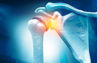
از عمل آرتروسکوپی میتوان برای تشخیص و درمان اختلالات مختلف مچ پا استفاده کرد. لیست مشکلاتی که میتوان آنها را به کمک آرتروسکوپی درمان کرد، روزبهروز در حال تکامل بوده و موارد زیر در آن قرار دارند
آرتروز مچ پا: عمل خشک کردن مفصل مچ پا برای بسیاری از بیماران مبتلا به شدیدترین حالت آرتروز مچ پا مناسب به نظر میرسد. به وسیلهی آرتروسکوپی میتوان عمل فیوژن یا خشککردن را بهصورت کم تهاجمی انجام داد. نتایج این عمل با عمل جراحی باز برابری کرده و یا حتی بهتر از آن خواهند بود
شکستگیهای مچ پا: عمل آرتروسکوپی را میتوان همراه با تکنیکهای جراحی باز به کار برد. این کار بهمنظور اطمینان از قرارگیری صحیح استخوانها و غضروفها در محل خود انجام میشود. همچنین میتوان از این عمل برای مشاهدهی آسیب غضروفها در طول عملترمیم مچ پا استفاده کرد
ناپایداری مچ پا: رباطهای مچ پا ممکن است دچار کشیدگی شده و باعث احساس بیثباتی در مفصل گردند. میتوان به کمک عمل جراحی، این رباطها را دوباره سفت کرد. تکنیکهای آرتروسکوپی میتوانند در حل این مشکل کارساز باشند
گیر افتادگی قدامی مچ پا (با نامهای مچ پای ورزشکاران و مچ پای فوتبالیستها نیز شناخته میشود): گیر افتادگی مچ پا زمانی اتفاق میافتد که استخوان و بافتهای نرم در جلوی مفصل مچ پا دچار التهاب شوند. از علائم این عارضه میتوان به درد و ورم مچ پا اشاره کرد. این امر موجب محدودیت مچ پا برای خم شدن رو به بالا خواهد شد. راه رفتن به سمت بالای شیب معمولا برای افراد مبتلا به این عارضه دردناک خواهد بود. استئوفیت (خارهای استخوانی) به وسیلهی عکسبرداری با اشعهی ایکس تشخیص داده میشوند
فیبروز مفصلی: بافت زخم (اسکار) ممکن است در فضای مفصلی مچ پا رشد کند. این امر موجب گرفتگی و درد در مفصل خواهد شد. به این عارضه فیبروز مفصلی یا آرتروفیبروز گفته میشود. میتوان از عمل آرتروسکوپی برای تشخیص محل ایجاد بافت زخم و برداشتن آن استفاده کرد
عفونت: عفونت فضای مفصلی را نمیتوان تنها به وسیلهی داروهای آنتی بیوتیک از بین برد. برای درمان عفونت معمولا یک عمل جراحی فوری برای شستشوی مفصل انجام میشود. میتوان این عمل را با روش آرتروسکوپی انجام داد
اجسام شناور: غضروف، استخوان و بافت اسکار ممکن است در فضای مفصلی رهاشده و اجسام شناور را به وجود بیاورند. این اجسام میتوانند موجب ایجاد درد و مشکلاتی چون صداهای غیرطبیعی مفصل و گیرکردن آن بشوند. قفل شدن مفصل مچ پا ممکن است اتفاق بیفتد. میتوان از عمل آرتروسکوپی مچ پا برای پیدا کردن و از بین بردن این اجسام استفاده نمود
نقصهای استئوکندرال (OCD): مناطقی از مفصل مچ پا که غضروف و استخوان آنها آسیبدیده است. این نقوص معمولا در اثر آسیب به مچ پا مانند پیچخوردگی و شکستگی ایجاد میشوند. از علائم شایع آن میتوان به درد و ورم در مچ پا اشاره کرد. بیماران ممکن است صداهای غیر طبیعی از داخل مفصل را شنیده و یا گیر افتادن آن را تجربه کنند. تشخیص این عارضه به کمک ترکیبی از معاینهی فیزیکی و عکسبرداری انجام میشود
گیر افتادگی خلفی مچ پا: این عارضه زمانی اتفاق میافتد که بافتهای نرم در پشت مچ پا دچار التهاب شده باشند. در طی این عارضه خم کردن پا به سمت پایین با درد همراه خواهد بود. این عارضه که در اثر استفادهی بیش از حد مفصل ایجاد میشود، در بین رقاصها رواج دارد
سینوویت: پوشش بافت نرم مفصل مچ پا (بافت سینوویال) ممکن است دچار التهاب شود. این عارضه باعث ایجاد درد و تورم شده و ممکن است در اثر آسیب و یا استفادهی بیش از حد ایجاد شده باشد. آرتروز التهابی (آرتریت روماتوئید) و استئوآرتریت نیز میتوانند موجب بروز سینوویت شوند. آرتروسکوپی مچ پا میتواند برای برداشتن بافت ملتهب شده و درمان مواردی که به روشهای درمانی بدون جراحی پاسخی ندادهاند استفاده شود
علائم غیر قابل توضیح در مچ پا: در برخی از موارد، بیماران علائمی را تجربه میکنند که نمیتوان آنها را با کمک تکنیکهای تشخیصی توضیح داد. آرتروسکوپی این فرصت را فراهم میکند تا به طور مستقیم، درون مفصل را مشاهده کرد. سپس جراح میتواند مشکلات احتمالی را شناسایی کرده و آنها را با کمک عمل جراحی رفع کند
کف پای صاف عارضه ای است که در آن بیماران فاقد قوس های استاندارد درکف پا هستند. قوس های موجود در کف پا نیروهایی که از طرف زمین به بدن اعمال میشود را کاهش داده و اجازه ورود به همه نیروهای وارده را به بدن نمی دهند اما در افراد با کف پای صاف،میزان زیادی این نیروها به دلیل فقدان قوس پا به سمت بدن اعمال شده و در دراز مدت میتواند منجر به بروز عوارض زنجیره وار در تمام مفاصل بدن خصوصاً ستون فقرات می گردد
مشکل صافی کف پا، عموماً مادرزادی بوده و به نام نرمی مفاصل نیز معروف است. نرمی مفصل یا ligamentum laxity در ناحیه کف پا سبب میشود که قوس کف پا در هنگام ایستادن بر روی پا از بین برود؛ همچنین تشکیل نشدن کامل یک یا دو استخوان در ناحیه مچ و کف پا و یا چسبیدن آنها به یکدیگر به صورت مادرزادی نیز در تشکیل نشدن قوس کف پا مؤثر است
اکثر کودکان بین ۱ تا ۵ سال دچار صافی کف پا هستند،که البته این بخشی از فرایند رشد طبیعی پاهای آنها است، و بیش از ۹۵ %از این کودکان در حین رشد صاحب قوس کف پایی طبیعی میشوند. اما ۵ %دیگر همچنان به صافی کف پا مبتلا خواهند بود. در اکثر موارد علت صافی کف پا، سست بودن اتصالات مفصلی میان استخوانهای مچ پا است. در این وضعیت رباط هایی که استخوانها را در کنار هم نگه میدارند شل بوده و هنگامی که کودک وزنش روی پایش قرار میدهد کشیده میشوند. همزمان که آنها رشد کرده و شروع به راه رفتن میکنند، بافتهای نرم موجود در کف پایشان هم سفت میشوند، که این باعث میشود قوس کف پا به تدریج شکل بگیرد
هنگامی که یک کودک مبتلا به صافی کف پا میایستد، قوس کف پایش ناپدید میشود. اما در هنگام نشستن یا زمانی که کودک روی نوک پنجه هایش میایستد، قوس کف پا دوباره ظاهر میشود. معمولا والدین یا سایر اعضا خانواده نگران این هستند که مبادا پایین بودن غیرطبیعی ارتفاع قوس کف پای کودک یا عدم وجود آن منجر به ایجاد بدشکلی یا معلولیت های دائمی در او شود، که البته در اغلب موارد این نگرانیها بی مورد است
هالوکس والگوس یا همان بونیون پا یک برجستگی استخوانی است ،که از تعویض مجدد مفصل در پایه انگشت بزرگ پا ایجاد می شود ؛ هالوکس والگوس بونیون شست پا اغلب بر قسمت درونی پا در مرکز انگشت شست پا در پایه پا تاثیر می گذارد، و همچنین می تواند بر خارج انگشت شست نیز تاثیر گذار باشد این موضوع، درزنان و مردان رخ می دهد اما در زنان بیشتر است
این عارضه،گاهی با علائمی همراه است، و گاه بدون علائم می باشد عارضه ای که یک تغییر شکل پیشرونده است و در یک جمله کوتاه راههای درمان آن می تواند شامل استراحت، سرمازدگی، تغییر کفش، پشتیبان پا (ارتوپیک)، داروها، تزریق استروئید و یا جراحی باشد
در حالی که علت دقیق شناخته نشده است، اعتقاد بر این است که به علت عوامل متعددی از جمله عملکرد غیرطبیعی پا و مکانیک، از قبیل ، آناتومی غیرطبیعی در ابتدا MTP مشترک و عوامل ژنتیکی ایجاد می شود. عوامل شایع در به وجود آمدن باعث شده که این مورد در جوانان بیشتر باشد ؛ بیومکانیک غیر طبیعی می تواند منجر به بی ثباتی ناشی از اختلال فالانگال متاتارس و اختلالات عضلانی شود که موجب تغییر شکل پذیری می شود
آناتومی غیرطبیعی، در اولین MTP مفصل می تواند یک شخص را در معرض انحطاط پینه قرار دهد ؛ یک مطالعه نشان می دهد ، که یک وراثت پذیری ژنتیک قابل توجهی از ناهنجاری های پینه ای در بین نژاد قفقاز اروپایی وجود دارد
هرچند کفش به طور مستقیم باعث ایجاد پینه و برآمدگی نمی شود، ولی مطمئنا وضعیت را درد آور و متورم تر می کند ؛ دیگر عواملی که کمتر شایع هستند از این نوع ناهنجاری، شامل آسیب هایی به (کشش، شکستگی و آسیب عصبی)، اختلالات عصبی یا عضلانی (بیماری فلج اطفال یا بیماری قلبی- امراض دندان) و اختلاف طول اندام (یک پا کوتاهتر از دیگر هستند). عضو بلندتر، باعث خم و چرخش بیشتر می شود
Arthroscopy
Arthroscopy (also called arthroscopic or keyhole surgery) is a surgical procedure that can be used to diagnose and treat various ankle disorders. The list of disorders treatable with arthroscopy is getting longer day-by-day; however, some of them are as follows:
Ankle Arthritis
The procedure of artificial ankylosis (also called arthrodesis or syndesis) is appropriate for most patients with the most severe forms of ankle arthritis. Using arthroscopic surgery, fusion can be done less invasively. The results of this operation will be equal to or even better than that of open surgery.
Ankle Fractures
Arthroscopic surgery can be used with open surgery techniques. They do this to ensure that the bones and cartilages are positioned properly. Moreover, doctors use arthroscopic surgery to observe cartilage damage during ankle repair surgery.
Ankle Instability
Ankle ligaments may be torn and cause instability in the joint. Surgery can restore these ligaments to their previous natural condition. Arthroscopic techniques can help solving ankle instability.
Anterior Ankle Impingement
An athletes’ disease, it occurs when the bones and soft tissues of the anterior part of the ankle joint are inflamed. Symptoms include ankle pain and swelling. This condition can cause limitations in the ankle for bending upward. Walking up the slope will usually be painful for people with this condition. Osteophytes are diagnosed by X-ray imaging.
Arthrofibrosis
Scar tissue may grow in the ankle joint space. This will cause cramps and pain in the joint. This is called arthrofibrosis. Arthroscopic surgery can be used to diagnose the place the scar tissue is formed and to remove it.
Infection
Antibiotics alone cannot eliminate joint space infection. To treat infection, they usually perform an urgent surgery. This surgery can be done with arthroscopy.
Floating Objects
Cartilage, bone and scar tissue may freely float inside joint space and form floating object. These objects can cause pain and problems such as abnormal joint sounds and cracking. Moreover, locks of the ankle joint can also occur. Nevertheless, you should not worry because ankle arthroscopy can be used to find and eliminate these objects.
Osteochondrial Defects (OCD)
Parts of the ankle joint that their cartilage and bone have been injured. These often occur due to ankle injuries like sprained or fractured ankle. Common symptoms include pain and swelling in the ankle. Patients may hear the joints sounding off or experience their impingement. The diagnosis is done by a combination of physical examination and imaging.
Posterior Ankle Impingement
This condition occurs when the soft tissues at the posterior part of the ankle are inflamed. In this condition, bending the feet downward would be painful. Posterior ankle impingement is more common among dancers due to overuse of the joints.
Synovitis
Soft tissue covering of the ankle joint (synovial tissue) may inflame. Synovitis may be caused by injury or overuse and can cause pain and swelling. Rheumatoid arthritis and osteoarthritis can also cause synovitis. Ankle arthroscopy can be used to remove the inflamed tissue and to treat cases that have not been treated using non-surgical treatment procedures.
Unexplainable Ankle Symptoms
In some cases, patients experience symptoms that cannot be explained by diagnostic techniques. Arthroscopy provides the opportunity to see directly within the joint. The surgeon can then diagnose any potential problems and solve them using surgery.
Flat feet is a condition in which patients have their foot arches collapsed and the whole foot sole comes into complete or near-complete contact with the ground. The arches on the soles of the foot reduce the forces imposed on the body from the ground and do not allow the forces to be imposed on the body completely. However, in people with flat feet, great amounts of these forces to the body make difficulties to them. In the long-term, it can lead to continuous side effects on all the joints of the body, especially the spine.
The flat feet disorder is generally congenital and is known as ligamentous laxity. Ligamentous laxity in the soles of the foot causes the arch of the soles to disappear while standing. In addition, in not forming the arch of the foot, incomplete formation of one or two bones in the ankle and foot or congenital adhesions of them are also effective.
Most of the children between 1 and 5 years old suffer from flat feet. This is part of their natural feet growth, 95% of these children will grow natural arches in their soles, and the rest 5% will grow flat foot. In most cases, the cause of flat foot is loose joint between anklebones. In this condition, the ligaments that hold the bones together become loose and sprained when the baby puts his/her weight on his/her foot. As they grow and begin to walk, the soft tissues in the soles of their feet also tighten, causing the arch of the sole to form gradually.
When a child with flat foot stands, the sole arches of his/her feet disappear. However, when sitting or standing on the tip of his/her toes, the arch of the soles of the foot reappears to the vision. Parents or other family members often worry that the abnormal low distance between the children’s arches sole to the ground or having no arches will lead to permanent malformations or disabilities. In most cases, however, you need not to worry at all.
Hallux valgus (also called Bunion) is a bony protrusion, caused by joint replacement at the base of the big toe. Hallux valgus of the big toe often affects the inner part of the feet in the center of the big toe at the base of the foot. It can also affect the outer part of the big toe, as well. It occurs in both men and women but is more common in women. It sometimes shows its symptoms, and sometimes not. This is a progressive deformity and its treatment ways can include resting, changing your shoe size, using orthopedic insoles, taking medicines, injecting steroids or undergoing surgery.
While the exact cause is unknown, many believe that it is caused due to several factors including abnormal foot function and mechanics, like metatarsophalangeal joints (MTP joints) and genetic factors. This mostly occurs in young adults. Abnormal biomechanics can lead to instability caused by metatarsal phalanx disorder and muscle disorders that cause deformability.
The abnormal anatomy in the first MTP joints can expose a person to callus removal. One study shows that there is a significant genetic heritability of callus abnormalities among Caucasian Europeans.
Although shoes do not directly cause calluses or bumps, they certainly make the condition more painful and swollen. Other less common causes of this type of anomaly include injuries (sprain, fracture and nerve damage), neurological or muscular disorders (poliomyelitis, heart and dental diseases) and limb length discrepancy (one foot shorter than the other). The longer the organ, the more it will bend and rotate.